COW-2020-24

CASE OF THE WEEK

2020-24 / June 15
(Contributor: Daniel Athanazio)

A 60-year old patient underwent a transurethral resection of the prostate due to clinical diagnosis of nodular hyperplasia.

Quiz

1. What is the diagnosis?

a. Metastatic melanoma to the prostate

b. Blue nevus of the prostate

c. Old (chronic) hemorrhages in prostatic stroma

d. Prostatic melanosis

e. Pigmented schwannoma

1. Blue nevus of the prostate

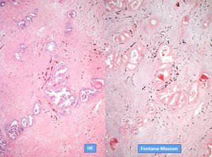
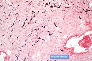
Lesions containing melanin in the prostate are classified as melanosis, blue nevus, and primary/metastatic malignant melanoma. The presence of heavily pigmented dendritic melanocytes characterizes blue nevus. The term melanosis implies that melanin pigment is observed in the glandular cells and in the stroma. Both lesions are rare with about 30 cases reported for each of them in English literature. They are almost always an incidental finding of TURP specimens.

Differential diagnosis with melanoma relies in the presence of obvious hypercellulary, cytologic atypia and mitotic activity in the latter. No features of malignancy were seen in the present case. In addition, no pigment within epithelial cells was detected.

1. Paner GP, Aron M, Hansel DE, Amin MB. Non-epithelial neoplasms of the prostate. Histopathology. 2012;60:166–86.

2. Gucer H, Bagci P. Prostatic melanosis. Urol Ann. 2014 Oct;6(4):384-6.

3. Farran EJ, Kerr PS, Kosarek CD, Sonstein J, Eyzaguirre EJ. A Rare Case of Multifocal Prostatic Blue Nevus. Case Rep Urol. 2018 Feb 21;2018:7820717.

Daniel Athanazio
Professor of Medicine, Federal University of Bahia
Imagepat, Laboratory of Pathology
Salvador, Bahia, Brazil

Prostate

Prostate; Melanosis; Blue nevus