

CASE OF THE WEEK
2021-15/ April 12
Contributors: Katherine Saunders and Sara E. Wobker
A man in his 40s presented with right testicular pain. He underwent right orchiectomy, which showed a 1.1 cm tumor centered in the mediastinum testis.


Quiz
1. What is the correct diagnosis?
a. Tubular ectasia of the rete testis
b. Sertoliform cystadenoma of the rete testis
c. Papillary cystadenoma of the epididymis
d. Rete testis adenocarcinoma
e. Adenomatous hyperplasia of the rete testis
1. b
1. Sertoliform cystadenoma of the rete testis
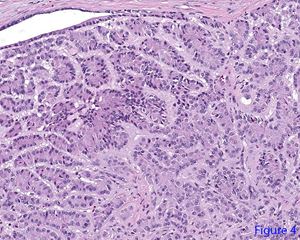
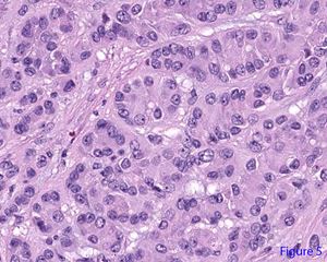
Sertoliform cystadenomas of the rete testis are extremely rare with very few cases reported in the literature. These tumors are benign and usually cured with orchiectomy. At low power, the lesion appears confined to the rete testis with predominantly intra-rete growth (figure 1). It is composed of lobules separated by fibrous stroma (figure 2), with non-destructive extension of the tumor cells into a hyaline and myxoid stroma (figure 3). Within the lobules, the tumor cells are arranged in tubules, cords, and nests (figure 4). The tumor cells are round to oval with open chromatin, prominent nucleoli, and eosinophilic cytoplasm (figure 5). The cells are mildly pleomorphic with no appreciable mitotic activity. The tumor expressed calretinin, inhibin, and vimentin. It lacked expression of SALL-4 and synaptophysin. A Ki-67 proliferation index stained less than 1% of the cells.
Tubular ectasia of the rete testis is a benign entity characterized by cystic dilatation of the rete testis due to obliteration of the efferent ducts. A papillary cystadenoma of the epididymis is another rare, benign neoplasm associated with von Hippel-Lindau syndrome. This entity shows cysts containing papillary projections lined by bland cuboidal cells with clear cytoplasm, similar in appearance to clear cell papillary renal cell carcinoma. Rete testis adenocarcinoma is a rare, malignant neoplasm usually occurring in older men. The tumor usually has a papillary, tubulopapillary, or glandular architecture, and an important diagnostic clue is finding the transition from normal rete testis to tumor with destructive invasive growth pattern. In adenocarcinoma, there is moderate to marked cellular pleomorphism and often necrosis is present. Adenomatous hyperplasia is usually a small, incidental finding characterized by a bland, tubulopapillary proliferation which projects into the channels of the rete testis, which may occur in association with germ cell tumors.
1. Paluru S; Ulbright T; Amin M; Montironi R; Epstein JI. The Morphologic Spectrum of Sertoliform Cystadenoma of the Rete Testis. Am J Surg Pathol. 2018;42(2):141-149.
2. Al-Obaidy KI, Idrees MT, Grignon DJ, Ulbright TM. Adenocarcinoma of the Rete Testis: Clinicopathologic and Immunohistochemical Characterization of 6 Cases and Review of the Literature. Am J Surg Pathol. 2019;43:670-681.
3. Odrzywolski KJ, Mukhopadhyay S. Papillary cystadenoma of the epididymis. Arch Pathol Lab Med. (1976). 2010;134:630-633.
Katherine Saunders
Sara E. Wobker
University of North Carolina School of Medicine
Chapel Hill, North Carolina
Testis
Rete testis, sertoliform cystadenoma, paratestis